Type
Presentation
Event
EPPC (Padua, Italy)
Authors
Spencer ART, Kenrick P, Steart DC, Garwood RJ, Hilton J, Munt MC, Needham J
Date
2014
Abstract
The Cheirolepidiaceae are an extinct group of conifers with an extensive Mesozoic fossil record well-documented by foliage, wood, pollen and reproductive organs. Three-dimensionally preserved Cheirolepidiaceous ovuliferous cones are rare, with few known species. Here we use X-Ray computed micro-tomography (XMT) at the Diamond Light Source Synchrotron (Oxfordshire, UK) to non-destructively image the internal anatomy of a recently discovered petrified ovuliferous cone from a Jurassic (Tithonian Stage: ca 145 million years) forest in southern England. The cone is derived from a suite of silicified plant materials, most of which were permineralised in situ in a hyper saline lagoonal setting during a shallow transgressive phase.
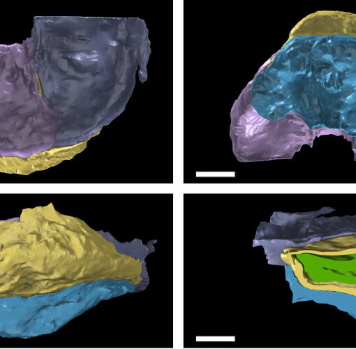
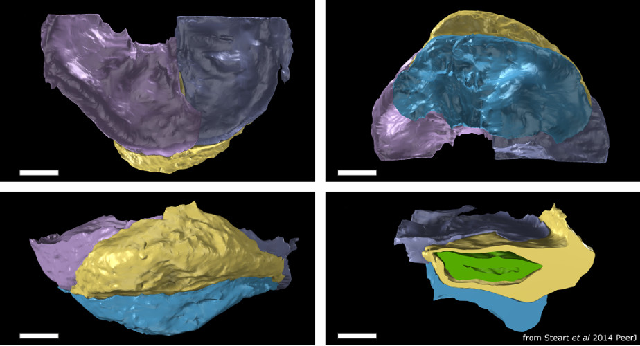
The petrified cone is small (1.2 x 1.3 cm) and unique, so we chose to employ non-destructive visualization techniques to reconstruct key anatomical characters. We used the Diamond I12 – JEEP (Joint Engineering, Environment and Processing) beamline, which was selected for its high brilliance and ability to generate high resolution (< 5µm) tomographic scans on macro-sized (< 25mm) objects. Tomographic datasets were used to fully describe the histology of the cone and produce three-dimensional models of the gross morphology using SPIERS and Blender™.
The tomographic images revealed a remarkable degree of information on the internal tissues of the cone, resolving cellular level details. We were able to identify and characterize bracts, ovuliferous scales, ovules and cellular features within. The form and patterning of vascular tissues was also discernible. Using SPIERS software, distinct anatomical structures were assigned to individual ‘masks’ in order to be rendered as separate isosurfaces. This process, improved through iterative editing and rendering, allowed the production of precise false colour models displaying well defined anatomical features within three-dimensional space. The bract/scale complexes were shown to have large ovuliferous scales, subtended by broad bracts, each containing only one ovule. A total of 14 ovules were identified. Each ovule was inverted and enclosed by pocket-forming tissue, opening at the micropyle proximal to the cone axis. The bracts and scales are fed by separate vascular traces, with the bract-trace diverging distally into multiple smaller bundles and the scale-trace forming a vascular pad for the ovule.
Our results demonstrate that the cone belongs to the extinct genus Pararaucaria (Cheirolepidiaceae) that has only previously been reported from South and North America, and forms only the fourth member of the genus to be described. This new species extends the known geographic range of Pararaucaria to the Northern edge of the newly forming Atlantic Ocean and emphasises the wide distribution of Pararaucarian conifers during the Jurassic.
Download (.pdf): To be added soon…
Slides
To be added soon…
