Type
Presentation
Event
The Linnean Society of London: Palaeobotany Specialist Group Meeting
Authors
Spencer ART, Sutton MD
Date
2010
Abstract
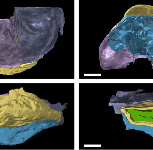
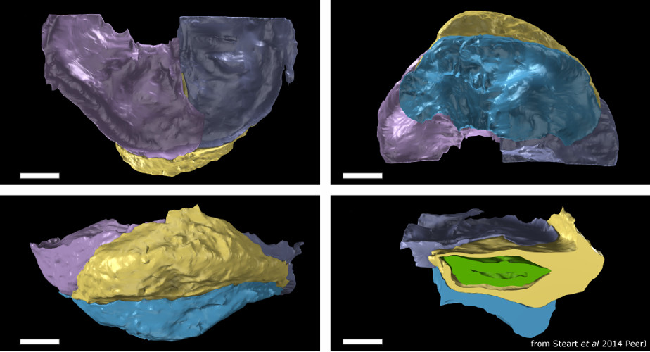
Over the last decade Micro-CT or X-ray Micro Tomography (XMT), has become increasingly available as an investigative tool. Development of scanning technology has been matched by a rise in computational power and advances in software, reducing the financial and time costs associated with the process. Three-dimensional reconstruction via XMT can hence now move from being reserved for one-off samples with known preservation to a preliminary method of investigation. Palaeobotanical material is often three-dimensionally preserved, and hence well suited to tomographic reconstruction. XMT-reconstruction requires X-ray density contrast between specimen and matrix; while this is present in some plant fossils, which scan well, those preserved as organic material can be ‘difficult’. Nonetheless even in these cases XMT data can provide preliminary 3D reconstructions which can serve as an important guide for more traditional (destructive) investigative techniques, such as serial grinding, enabling them to capture the maximum amount of data. This preliminary-scan approach is especially important where anatomically preserved organs which are hitherto unknown are to be investigated. We report here the preliminary results from a project to investigate the anatomy and affinity of a range of Palaeozoic plant-organ fossils using XMT.
Download (.pdf): LinSoc 2010 PDF
Slides


