Type:
Peer Reviewed Research Paper
Journal:
PeerJ
Year:
August 28 2017
Authors:
Alan R.T. Spencer, Russell J. Garwood, Andrew R. Rees, Robert J. Raine, Gar W. Rothwell, Neville T.J. Hollingworth, Jason Hilton
DOI
https://doi.org/10.7717/peerj.3723
Abstract:
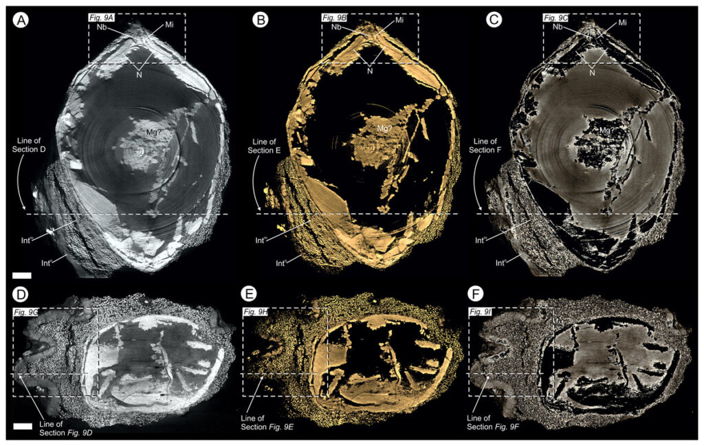
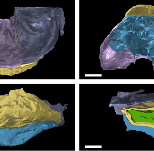
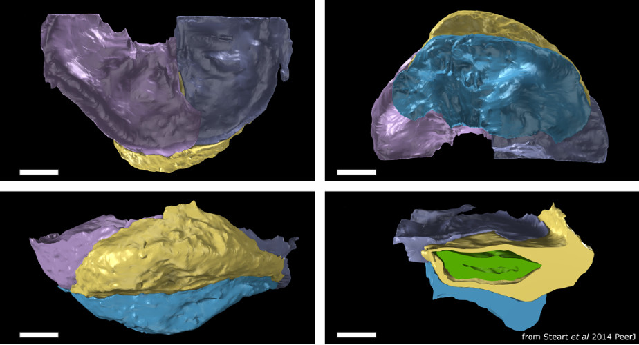
Most knowledge concerning Mesozoic Era floras has come from compression fossils. This has been augmented in the last 20 years by rarer permineralized material showing cellular preservation. Here, we describe a new genus of anatomically preserved gymnosperm seed from the Callovian–Oxfordian (Jurassic) Oxford Clay Formation (UK), using a combination of traditional sectioning and synchrotron radiation X-ray micro-tomography (SRXMT). Oxfordiana motturii gen. et sp. nov. is large and bilaterally symmetrical. It has prominent external ribs, and has a three-layered integument comprising: a narrow outer layer of thick walled cells; a thick middle parenchymatous layer; and innermost a thin fleshy layer. The integument has a longitudinal interior groove and micropyle, enveloping a nucellus with a small pollen chamber. The large size, bilateral symmetry and integumentary groove demonstrate an affinity for the new species within the cycads. Moreover, the internal groove in extant taxa is an autapomorphy of the genus Cycas, where it facilitates seed germination. Based upon the unique seed germination mechanism shared with living species of the Cycadaceae, we conclude that O. motturii is a member of the stem-group lineage leading to Cycas after the Jurassic divergence of the Cycadaceae from other extant cycads. SRXMT—for the first time successfully applied to fossils already prepared as slides—reveals the distribution of different mineral phases within the fossil, and allows us to evaluate the taphonomy of Oxfordiana. An early pyrite phase replicates the external surfaces of individual cells, a later carbonate component infilling void spaces. The resulting taphonomic model suggests that the relatively small size of the fossils was key to their exceptional preservation, concentrating sulfate-reducing bacteria in a locally closed microenvironment and thus facilitating soft-tissue permineralization.
Additional SI:
The following information was supplied regarding data availability:
Zenodo–https://zenodo.org
Video S1: https://doi.org/10.5281/zenodo.61841
Video S2: https://doi.org/10.5281/zenodo.61841
Video S3: https://doi.org/10.5281/zenodo.61841
Model S1: https://doi.org/10.5281/zenodo.61841
Model S2: https://doi.org/10.5281/zenodo.61841
Figure S1: https://doi.org/10.5281/zenodo.61841
Dataset S1: https://doi.org/10.5281/zenodo.824099
Dataset S2: https://doi.org/10.5281/zenodo.824103
Dataset S3: https://doi.org/10.5281/zenodo.824029
Dataset S4: https://doi.org/10.5281/zenodo.824047
Dataset S5: https://doi.org/10.5281/zenodo.824051
Dataset S6: https://doi.org/10.5281/zenodo.824073
Dataset S7: https://doi.org/10.5281/zenodo.824079
Dataset S8: https://doi.org/10.5281/zenodo.824085
Dataset S9: https://doi.org/10.5281/zenodo.824089
Dataset S10: https://doi.org/10.5281/zenodo.824091
