Type:
Peer Reviewed Research Paper
Journal:
Philosophical Transactions of the Royal Society B: Biological Science
Year:
December 18 2017. Volume published 5 February 2018.
Authors:
Christine Strullu-Derrien, Alan R. T. Spencer, Tomasz Goral, Jaclyn Dee, Rosmarie Honegger, Paul Kenrick, Joyce E. Longcore, Mary L. Berbee
DOI
http://dx.doi.org/10.1098/rstb.2016.0502
Abstract:
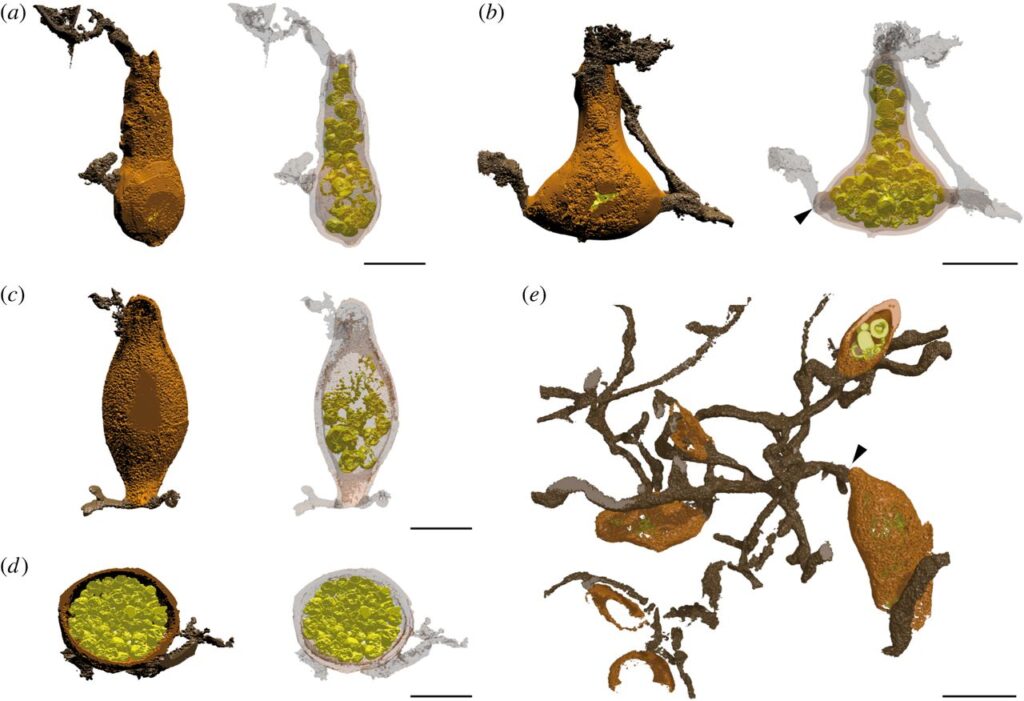
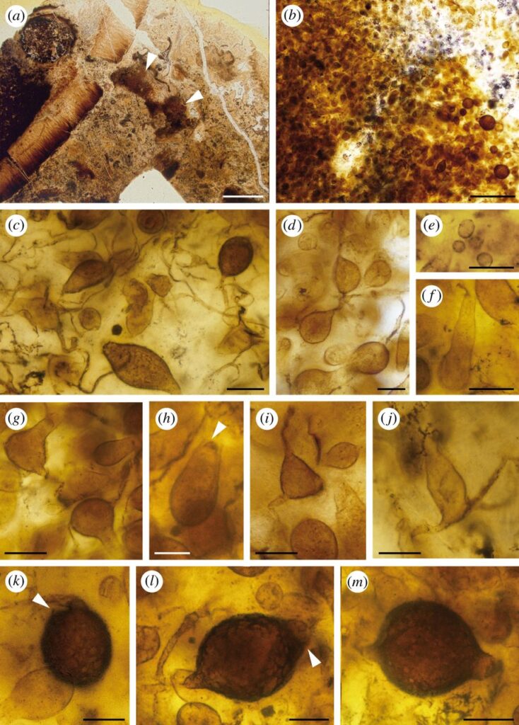
Zoosporic fungi are key saprotrophs and parasites of plants, animals and other fungi, playing important roles in ecosystems. They comprise at least three phyla, of which two, Chytridiomycota and Blastocladiomycota, developed a range of thallus morphologies including branching hyphae. Here we describe Retesporangicus lyonii gen. et sp. nov., an exceptionally well preserved fossil, which is the earliest known to produce multiple sporangia on an expanded hyphal network. To better characterize the fungus we develop a new method to render surfaces from image stacks generated by confocal laser scanning microscopy. Here, the method helps to reveal thallus structure. Comparisons with cultures of living species and character state reconstructions analysed against recent molecular phylogenies of 24 modern zoosporic fungi indicate an affinity with Blastocladiomycota. We argue that in zoosporic fungi, kinds of filaments such as hyphae, rhizoids and rhizomycelium are developmentally similar structures adapted for varied functions including nutrient absorption and anchorage. The fossil is the earliest known type to develop hyphae which likely served as a saprotrophic adaptation to patchy resource availability. Evidence from the Rhynie chert provides our earliest insights into the biology of fungi and their roles in the environment. It demonstrates that zoosporic fungi were already diverse in 407 million-year-old terrestrial ecosystems.
Additional SI:
https://dx.doi.org/10.6084/m9.figshare.c.3925837.
Supplementary three-dimensional model data are available at the Zenodo repository: https://dx.doi.org/10.5281/zenodo.801505
